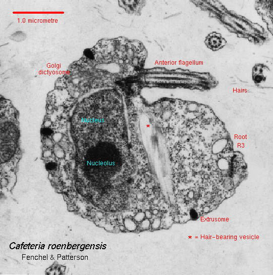
 The cells are uninucleate.
There is a single Golgi body anterior to the nucleus and near the
flagellar bases. About five
sausage-shaped mitochondria are present; as in other stramenopiles, the
cristae are tubular.
The cells are uninucleate.
There is a single Golgi body anterior to the nucleus and near the
flagellar bases. About five
sausage-shaped mitochondria are present; as in other stramenopiles, the
cristae are tubular.
As with most other stramenopiles, the anterior flagellum bears two rows of tripartite tubular hairs. The presence of three terminal fibrils, the central one longer than the two on the sides, helps distinguish Cafeteria from other naked stramenopile flagellates.
|
The cells are uninucleate.
There is a single Golgi body anterior to the nucleus and near the
flagellar bases. About five
sausage-shaped mitochondria are present; as in other stramenopiles, the
cristae are tubular. |
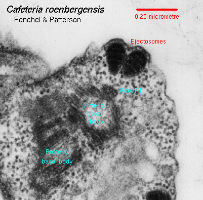 Ejectile organelles
(extrusomes) with a characteristic pattern dot the surface of the cell,
especially in the vicinity of the cytostome. Ejectile organelles
(extrusomes) with a characteristic pattern dot the surface of the cell,
especially in the vicinity of the cytostome. |
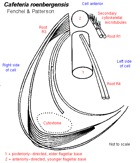 The cytoskeleton is
based on an asymmetrical system of two flagellar bases, three microtubular
roots, and a forked
rhizoplast. One of the three microtubular roots arises from the anterior
basal body, and has
secondary cytoskeletal microtubules associated with it; this is a common
feature among
stramenopiles. The other two microtubular roots arise from the posterior
basal body.
The cytoskeleton is
based on an asymmetrical system of two flagellar bases, three microtubular
roots, and a forked
rhizoplast. One of the three microtubular roots arises from the anterior
basal body, and has
secondary cytoskeletal microtubules associated with it; this is a common
feature among
stramenopiles. The other two microtubular roots arise from the posterior
basal body. |
 The broader (12
microtubules) of these two roots is subdivided distally into three
subunits that, together, define the feeding basket or cytostome. The broader (12
microtubules) of these two roots is subdivided distally into three
subunits that, together, define the feeding basket or cytostome. |
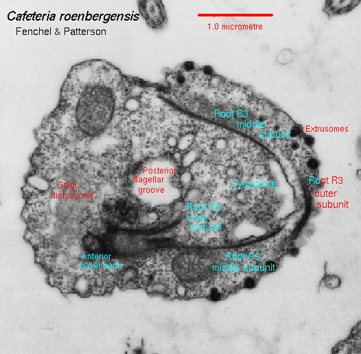 The
feeding basket is
displaced to the right of the axis defined by the posterior basal body and
flagellum. This feature,
plus the lack of an overlap between the distal ends of the two roots
arising from the posterior
basal body, help separate bicosoecids from other naked stramenopile
flagellates, especially the
colorless chrysophytes Paraphysomonas and Spumella. The
feeding basket is
displaced to the right of the axis defined by the posterior basal body and
flagellum. This feature,
plus the lack of an overlap between the distal ends of the two roots
arising from the posterior
basal body, help separate bicosoecids from other naked stramenopile
flagellates, especially the
colorless chrysophytes Paraphysomonas and Spumella.
|
Cafeteria: Index | Introduction | Appearance | Ultrastructure | Reproduction and Life History | Similar genera | Classification | Taxonomy and Nomenclature | Cultures | References | Internet resources
Protist Image Data: Picture Gallery | Home Page