Monosiga
Index
|
Introduction
|
Appearance
|
Ultrastructure
|
Reproduction and Life History
|
Similar genera
|
Classification
|
Taxonomy and Nomenclature
|
Cultures
|
References
|
Internet resources
ULTRASTRUCTURE
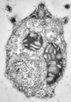
 Monosiga cells are
uninucleate (have one nucleus) and have one or more mitochondria,
dispersed throughout the cell. The mitochondrial cristae are flattened.
Unlike other genera of choanoflagellates in the family Monosigidae
(=Codosigidae), no theca is visible around the cells. At the
anterior end of the cell, a pair of basal bodies appears, only
one of which is associated with an emergent flagellum. An
electron-dense peripheral ring surrounds the flagellum-bearing
basal body. Beneath the basal body complex, a single Golgi
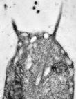
body occurs. Tentacles arise from various regions around the
cell and extend apically to form the collar. Food particles
are attached to the tentacles and transported to the cell body,
where they are digested in food vacuoles
|
Flagella are usually shown in light micrographs to lack any covering.
However, in at least one species, fine fibrils are present that form two
"paddles", one on either side of the flagellum.

|

|
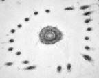
The cytoskeleton is based on a symmetrical system of
radially arranged
microtubules, and on tentacles (as well as other pseudopods that
may be produced at the base of the cell) that are supported by
microfilaments. The cytoskeletal microtubules arise from the
ring of electron-dense material that surrounds the
flagellum-associated basal body.
|
 The microfilaments supporting the
collar tentacles arise from the midregion of the cell.
|
The ultrastructural features of sessile cells and swarmers
(zoospores) are
similar, although tentacles on zoospores are usually shorter.
Monosiga: Index
|
Introduction
|
Appearance
|
Ultrastructure
|
Reproduction and Life History
|
Similar genera
|
Classification
|
Taxonomy and Nomenclature
|
Cultures
|
References
|
Internet resources
Protist Image
Data: Picture Gallery
|
Home Page
|
 The microfilaments supporting the
collar tentacles arise from the midregion of the cell.


