Monosiga
Monosigacells appear as sessile trophic cells and naked, free-swimming or creeping zoospores.
Left cell: sessile Right cell: zoospore |
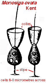 Stipitate species |
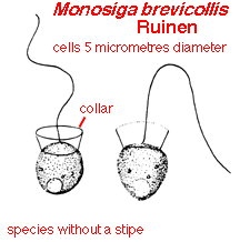
Trophic cells may be spherical to eggshaped to ellipsoidal (cucumber-shaped), depending on the species and/or growth conditions. A thin cell covering (theca) is usually only visible by electron microscopy except when a stipe is present. This feature helps separate Monosiga from its close relative Salpingoeca.
 DIC image | 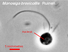 Phase contrast image |
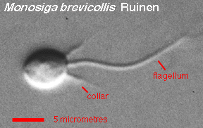
As is typical of choanoflagellates generally, the anterior end of the cell shows a single flagellum and a ring of tentacles. Under most light microscopes, the tentacle ring appears as a solid collar around the flagellum. Movement of the flagellum directs a current of water, containing potential food particles (including bacteria, the usual prey items) through the collar. Captured particles are transported down the tentacles of the collar to the cell body, where suitable ones are ingested. In disturbed cells, both the flagellum and the collar may be shed or retracted.
Zoospores are similar to the sessile cells but are highly motile and may not have an extended collar. Cells swim with the cell body forward, the flagellum trailing behind (like sperm cells of animals). During settlement, zoospores may produce fine pseudopodia at the posterior end of the cell, by which the cell slowly creeps to the position at which it will secrete its theca and become sessile.
Monosiga: Index | Introduction | Appearance | Ultrastructure | Reproduction and Life History | Similar genera | Classification | Taxonomy and Nomenclature | Cultures | References | Internet resources
Protist Image Data: Picture Gallery | Home Page