Paramoeba and Neoparamoeba species occur in marine environments. They are isolated from marine or estuarine waters and sediments, or appear as pathogens in marine fish and invertebrates. The only state observed is the trophont, which is a locomotory, floating, or sessile amoeba.
As with other "naked" amoebae ("gymnamoebae"; those amoebae that do not live within an obvious shell, or "test"), species-specific morphology only appears when the cell is attached to a substrate and is moving (the "locomotory" form of the amoeba).
|
|
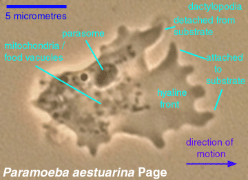
Paramoeba and Neoparamoeba amoebae in motion (picture at left; phase-contrast image) tend to be about twice as long as broad. Cell length varies among the species but typically ranges between 10 and 75 micrometres. The forward edge of the amoeba tends to be clear ("hyaline") and have several, more or less finger-like, shorter or longer, lobose pseudopods ("dactylopodia"). Some of the pseudopods are likely to be in contact with the substrate, while others will not be. The pseudopods, and the amoebae themselves, move detectably but slowly when viewed under the compound microscope at higher magnifications. |
|
Amoebae that are not in motion tend to lose the pseudopods and become more circular in outline. This is found most often in cells under stress, such as starvation in old cultures or coverslip pressure. Also, cells in tissues that are prepared for histological examination (killed, fixed, smeared or embedded in paraffin/resin and sectioned, and stained) usually do not preserve the morphology of the locomotive cell. |
Living amoebae that are detached from substrate express a "floating" form: approximately half a dozen fine unbranched pseudopodia radiating from a globular center. Nearly all gymnamoebae express such floating forms. It is very difficult, and not infrequently impossible, to distinguish one species from another on the basis of the morphology of the floating form.
|
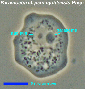
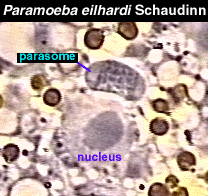
The cells are typically uninucleate, although occasional cells with two or more nuclei may be found. Adjacent to each nucleus is at least one parasome. The parasome is about the same size as the nucleus, but while the nucleus consists of nucleoplasm and a single centrally located nucleolus, the parasome consists of two small peripheral bodies and a central body. Often, the substructure of the parasome is not visible in living cells, appearing as a uniformly dark object in phase-contrast images. In histological preparations, the parasome stains with nucleophilic (e.g., hematoxylin) and DNA-specific (e.g. Feulgen) dyes. |
|
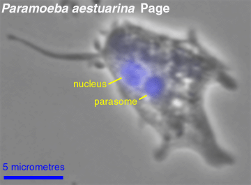
The parasome contains DNA. In the picture at left, a cell has been fixed and stained with the DNA-detecting fluorochrome dye DAPI (4',6-diaminidino-2-phenylindole). Under ultraviolet light, the complex formed between DNA and DAPI fluoresces blue. The color fluorescence image has been superimposed on a black-and-white phase-contrast image of the stained cell. |
In some studies, only the peripheral bodies of the parasome are said to stain with DNA-detecting dyes. In others, only the central region stains, and in still others, both peripheral and central bodies stain. Experience with DAPI suggests that DNA is present in both peripheral and central bodies, but that sometimes the parasome is so compact that the peripheral and central bodies cannot be distinguished easily. Also, the Feulgen procedure, the DNA-specific staining protocol used on parasomes (and other DNA-containing cell structures) before the DNA-detecting fluorochromes like DAPI came into general use, is tricky and does not always produce accurate or reliable results.
The parasome separates species of Paramoeba, Neoparamoeba and Janickina from all other amoebae. However, the absence of a a parasome is not necessarily informative, especially if, as seems likely, the parasome is an endocommensal protist related to bodonids that may be capable of independent dispersal. Some populations of P. eilhardi lack parasomes; these populations have been described as a separate species, Korotnevella nivo Smirnov (Smirnov 1996/97). Parasome-free populations of Neoparamoeba species have not yet been discovered.
To separate Paramoeba and Neoparamoeba from Janickina under the light microscope, one must see the number of pseudopods in the actively moving, attached amoeba. Janickina species have only one active pseudopod, Paramoeba and Neoparamoeba species have several. The character may not be observable in parasitic populations of Neoparamoeba.
Paramoeba: Index | Introduction | Appearance | Ultrastructure | Reproduction and Life History | Similar genera | Classification | Taxonomy and Nomenclature | Cultures | References | Internet resources
Protist Image Data: Picture Gallery | Home Page