 The first sign of imminent cell division is the formation of two
new basal
bodies in the vicinity of the old ones.
The first sign of imminent cell division is the formation of two
new basal
bodies in the vicinity of the old ones.
Reclinomonas cells reproduce asexually by binary division. No sexual reproduction has been observed.
|
The first sign of imminent cell division is the formation of two
new basal
bodies in the vicinity of the old ones.
|
 Just prior to
prophase, the basal bodies split into two pairs,
which then
migrate to opposite ends of the cell. Each pair consists of one "old" and
one "new" basal body. The "old" basal body in each pair will
become basal body 1 (eldest) in the progeny cells. Flagella remain
attached to the old basal bodies, and new flagella form on the new basal
bodies. Flagella remain attached throughout division. The microtubular
roots of the parent cell are severed from the separating basal body pairs
near their proximal ends (the ends nearest the basal bodies). The roots
then progressively depolymerize posteriorly.
Just prior to
prophase, the basal bodies split into two pairs,
which then
migrate to opposite ends of the cell. Each pair consists of one "old" and
one "new" basal body. The "old" basal body in each pair will
become basal body 1 (eldest) in the progeny cells. Flagella remain
attached to the old basal bodies, and new flagella form on the new basal
bodies. Flagella remain attached throughout division. The microtubular
roots of the parent cell are severed from the separating basal body pairs
near their proximal ends (the ends nearest the basal bodies). The roots
then progressively depolymerize posteriorly.
|
 |
 |
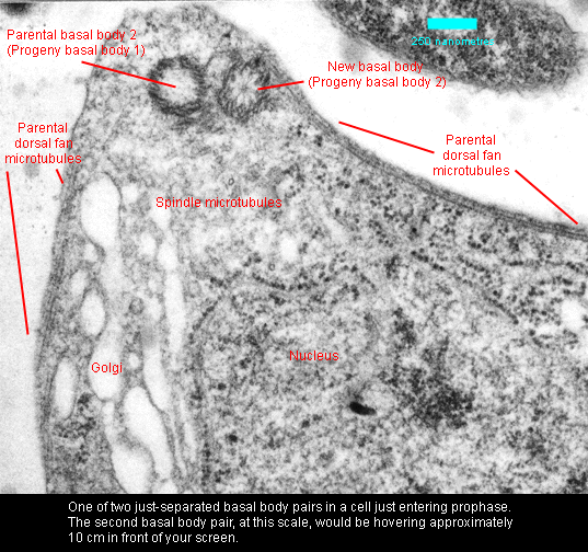
At prophase, the basal bodies are located at the spindle poles. Each basal body pair is the focus for a cone of microtubules that extends toward and envelops the nucleus. Also, at each basal body pair, new microtubular roots have begun to form. The nuclear envelope initially remains intact, but later dissipates, at which time the microtubules enter the nucleus and become associated with chromosomes. Chromosome condensation is evident at prophase. The nucleolus remains intact. |
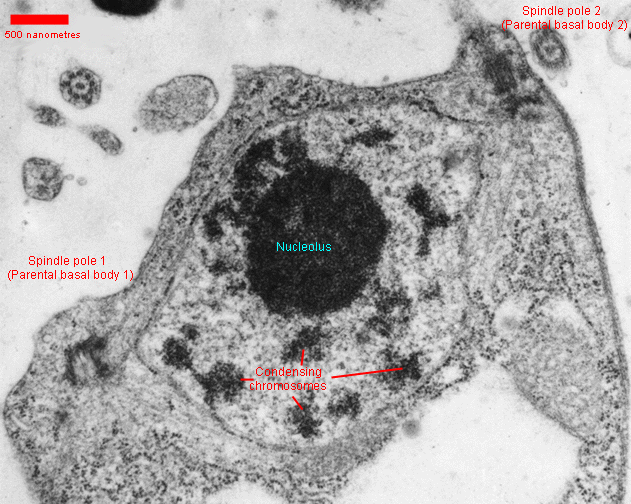
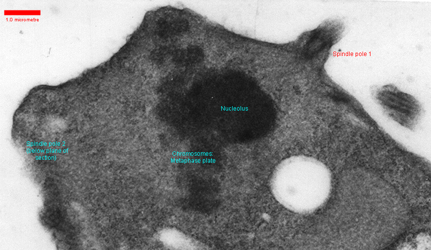 At metaphase, there
are two conical "half-spindles" focused on
the basal
body complexes and terminating at the chromosomal plate. Chromosomes are
small, irregularly shaped and difficult to count. The number appears to
be in th twenties or thirties. The nucleolus is persistent.
At metaphase, there
are two conical "half-spindles" focused on
the basal
body complexes and terminating at the chromosomal plate. Chromosomes are
small, irregularly shaped and difficult to count. The number appears to
be in th twenties or thirties. The nucleolus is persistent.
|
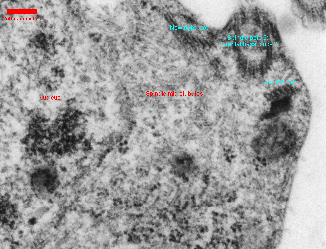
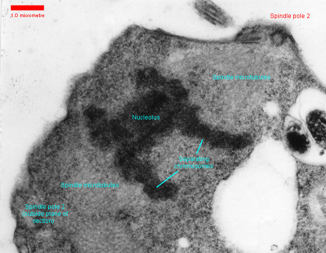 Chromosome movement at anaphase appears to be due mostly to the
separation of the spindle poles. There do not appear to be any
microtubules that extend from one spindle pole to the other (that is,
there is no "interzonal spindle"), and shortening of the microtubules in
the half spindles is minimal until late anaphase and telophase.
Chromosome movement at anaphase appears to be due mostly to the
separation of the spindle poles. There do not appear to be any
microtubules that extend from one spindle pole to the other (that is,
there is no "interzonal spindle"), and shortening of the microtubules in
the half spindles is minimal until late anaphase and telophase.
|
The progeny cell that receives the parent cell's anterior flagellum (basal body 2) also receives a large vesicle containing lorica scales. This cell becomes the zoospore and swims away. After an hour or so, this swimming cell settles, secreting its lorica, and its elder flagellum becomes associated with the ventral groove and develops a vane ... that is, the anterior flagellum of the parent cell becomes the posterior flagellum of the progeny cell.
The progeny cell that receives the parent cell's posterior flagellum remains in the parent lorica.
Return to summary information