Malawimonas
Ultrastructure
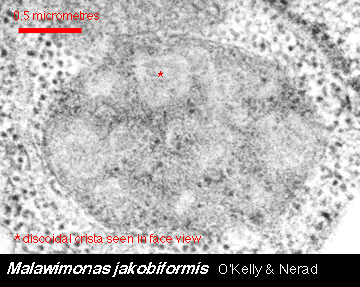
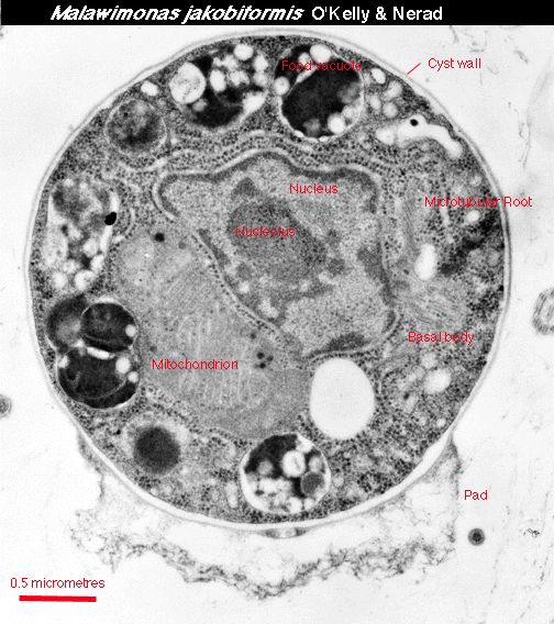
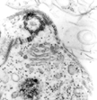
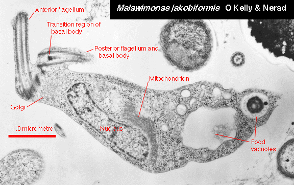
 A longitudinal section of the cell shows the anterior flagellar insertion
and the positions of major organelles. Food vacuoles contain
bacteria. The very short basal bodies, with transition regions
recessed well interior to the plane of the cell membrane, are
characteristic of Malawimonas and are not found in other
similar flagellates.
A longitudinal section of the cell shows the anterior flagellar insertion
and the positions of major organelles. Food vacuoles contain
bacteria. The very short basal bodies, with transition regions
recessed well interior to the plane of the cell membrane, are
characteristic of Malawimonas and are not found in other
similar flagellates. |
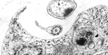
 The single mitochondrion is located next to, and mostly posterior to,
the nucleus. The cristae are discoidal.
The single mitochondrion is located next to, and mostly posterior to,
the nucleus. The cristae are discoidal. |
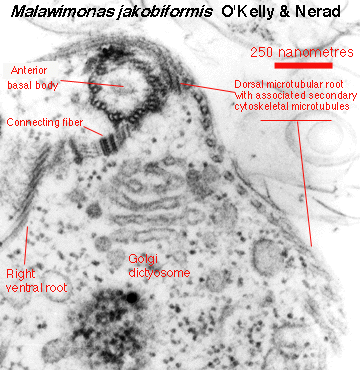
 A single Golgi body/dictyosome lies next to the flagellar
bases. This picture also shows some of the microtubular roots
associated with the anterior end of the cell (see the drawing of
the cytoskeleton, below).
A single Golgi body/dictyosome lies next to the flagellar
bases. This picture also shows some of the microtubular roots
associated with the anterior end of the cell (see the drawing of
the cytoskeleton, below). |
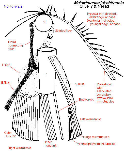
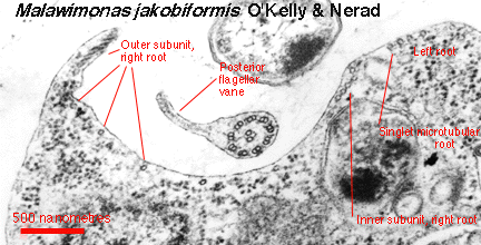
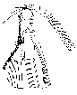 The cytoskeleton is an asymmetrical system of two flagellar bases,
three ventral microtubular roots, and a dorsal microtubular
root from which secondary microtubules arise and fan out over
the dorsal surface. Unlike other jakobids (Jakoba, Reclinomonas and
Histiona), the left ventral root is not associated with a multilayered
structure. Also, most of the microtubules that form the floor
of the ventral groove come from the right ventral root; in
other jakobids, these microtubules mostly come from the left
root.
The cytoskeleton is an asymmetrical system of two flagellar bases,
three ventral microtubular roots, and a dorsal microtubular
root from which secondary microtubules arise and fan out over
the dorsal surface. Unlike other jakobids (Jakoba, Reclinomonas and
Histiona), the left ventral root is not associated with a multilayered
structure. Also, most of the microtubules that form the floor
of the ventral groove come from the right ventral root; in
other jakobids, these microtubules mostly come from the left
root. |
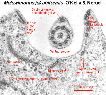
 The posterior
flagellum possesses a conspicuous and characteristic
flagellar vane. The left picture shows its origin, near the anterior end
of the cell. The right picture shows how the vane looks at approximately
the middle of the cell. This vane arises on the ventral
surface of the flagellum; in other
jakobids, the vane arises on the dorsal surface.
These pictures also show how the ventral microtubular roots appear in
different parts of the cell (compare the map of the cytoskeleton, above).
The posterior
flagellum possesses a conspicuous and characteristic
flagellar vane. The left picture shows its origin, near the anterior end
of the cell. The right picture shows how the vane looks at approximately
the middle of the cell. This vane arises on the ventral
surface of the flagellum; in other
jakobids, the vane arises on the dorsal surface.
These pictures also show how the ventral microtubular roots appear in
different parts of the cell (compare the map of the cytoskeleton, above).
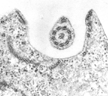
 The cyst is uninucleate, with one mitochondrion, and contains remnants of
the kinetid (basal bodies and
microtubular roots). It is surrounded by a thin wall, probably of organic
material. The pad consists of fibrillar material, possibly representing
adhesive polysaccharides. A pore in the wall in the vicinity of the pad
may represent an exit aperture.
The cyst is uninucleate, with one mitochondrion, and contains remnants of
the kinetid (basal bodies and
microtubular roots). It is surrounded by a thin wall, probably of organic
material. The pad consists of fibrillar material, possibly representing
adhesive polysaccharides. A pore in the wall in the vicinity of the pad
may represent an exit aperture. |
Return to summary information
 A longitudinal section of the cell shows the anterior flagellar insertion
and the positions of major organelles. Food vacuoles contain
bacteria. The very short basal bodies, with transition regions
recessed well interior to the plane of the cell membrane, are
characteristic of Malawimonas and are not found in other
similar flagellates.
A longitudinal section of the cell shows the anterior flagellar insertion
and the positions of major organelles. Food vacuoles contain
bacteria. The very short basal bodies, with transition regions
recessed well interior to the plane of the cell membrane, are
characteristic of Malawimonas and are not found in other
similar flagellates.