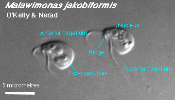
Malawimonas cells are encountered as naked freeswimming trophic cells and sessile cysts.
 |
Trophic cells are ellipsoidal to pear-shaped (the latter usually when the cells are compressed, as under a cover slip) and between 5 and 12 micrometers long. Two flagella emerge from the anterior end of the cell. The anterior flagellum is free; at rest, the flagellum often projects anteriorly at first and then "hooks back" ventrally. |
 |
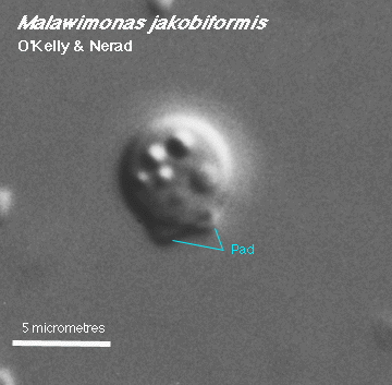
Cysts are formed from single trophic cells. They are spherical, with a thin wall probably composed of organic materials and lacking surface ornamentation. They have no aperture visible by light microscopy. They are attached to a substrate by a pad of material. |
Return to summary information