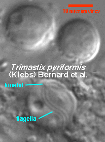
Differential interference contrast
Phase contrast
vanes
Trimastix cells are found in anoxic (no oxygen) or microaerobic (very low oxygen) environments as free-swimming trophic cells and (in one species) thin-walled cysts.
|
Differential interference contrast | Phase contrast | vanes |
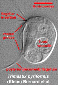
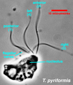
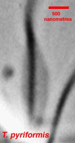
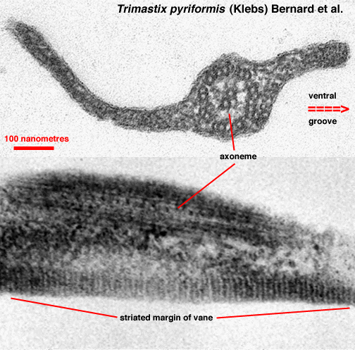
The trophic cells are more or less pear shaped. Depending on the species and on growth conditions, they may range from about 12 to about 25 micrometres in length. Four flagella emerge from just below the anterior end of the cell. Three of these flagella are "free", while the fourth (the posterior flagellum) normally remains in a groove on the ventral surface of the cell. The posterior flagellum may also be called the "recurrent" flagellum. Unlike "recurrent" flagella in some other protist groups, the posterior flagellum in Trimastix is not physically attached to the cell body, as can be seen in cells fixed for electron microscopy. In all Trimastix species, the posterior flagellum bears two vanes. In one species, T. marina, the anterior flagellum is conspicuously thicker than the rest. In other species, no special features are found on the non-posterior flagella.
Bacteria are captured in the ventral groove and are digested in food vacuoles.
Mitochondria are absent.
 |
Trimastix trophs closely resemble those of the
retortamonad protist
genus Chilomastix. Retortamonads also lack mitochondria. The single
nucleus in the Trimastix
trophic cell has a central nucleolus; this feature is absent
in species
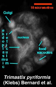
of Chilomastix. Trimastix cells also have Golgi and endoplasmic reticulum internal membrane systems. Both of these endomembrane systems may be observed either with transmission electron microscopy or with fluorescent labelling techniques. Here, cells have been incubated in the fluorescent lipid BODIPY FL C5 Ceramide, which is selectively taken up by Golgi and endoplasmic reticulum membranes (as well as by bacterial membranes in food vacuoles). The Golgi body lies near the flagellar bases, while the endoplasmic reticulum network mostly surrounds the nucleus. Chilomastix species lack endomembrane systems that can be seen by transmission electron microscopy or BODIPY Ceramide incubation. Other protists that resemble Trimastix, in particular the heterolobosean genera Tetramitus and Percolomonas, differ by lacking the vanes on the posterior flagellum. Most species assigned to Tetramitus and Percolomonas have mitochondria with discoidal cristae. |
 |
Cysts are known from a single culture strain of T.
pyriformis. The cysts are apparently formed
from single cells (i.e., not as a result of gamete fusion). They have
thin walls. Internally, they retain the flagella and kinetid of the parent
trophic cell. They are apparently short-lived; old cultures containing
them and retained at growth-permissive temperatures without medium change
or feeding with bacteria for more than a few weeks lose the ability to
produce trophic cells.
Chilomastix cysts are thick-walled and hardier, but otherwise closely resemble Trimastix cysts. Most other protists do not preserve flagella or kinetids in the cyst. |
Return to summary information