Chrysodidymus
ULTRASTRUCTURE

|
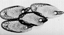 |
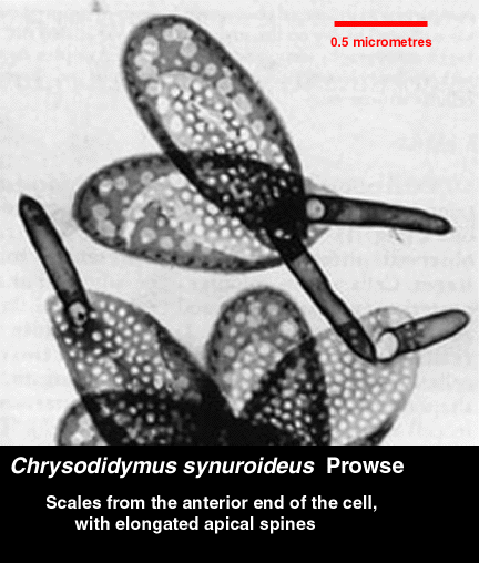
Chrysodidymus cell bodies are covered with a single layer of
silica scales. They are slipper-shaped with an apical spine. The
spine is longer on scales found at or near the anterior end of the
cell. |
 |
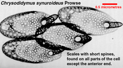
Scales are found on all parts of the cell body except
where the posterior ends of the cells adjoin. There are no obvious
structures that connect the two cells; presumably, an adhesive of
some sort is present that does not interact with electrons (and
therefore is invisible to electron microscopy).
|
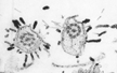 |
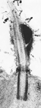
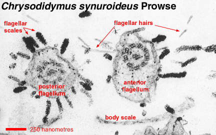
Scales also appear on both flagella. The longer, anterior flagellum
bears tripartite tubular hairs. These hairs are typical for members
of the stramenopile lineage.
|
|
The shorter, posteriorly-directed flagellum has a two-part flagellar
swelling near its proximal end (the "base" of the flagellum). This
flagellar swelling fluoresces green when illuminated with violet or
near-ultraviolet light. This autofluorescence is associated with
photoreceptor (light-absorbing) domains in golden algae and other protists
that are associated with phototaxis (the ability of cells to swim towards
or away from light).
The transition region of both flagella has a coiled fiber (transitional
helix) of six gyres. Many stramenopiles have transitional helices.
|
 |
 |
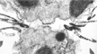
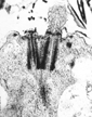
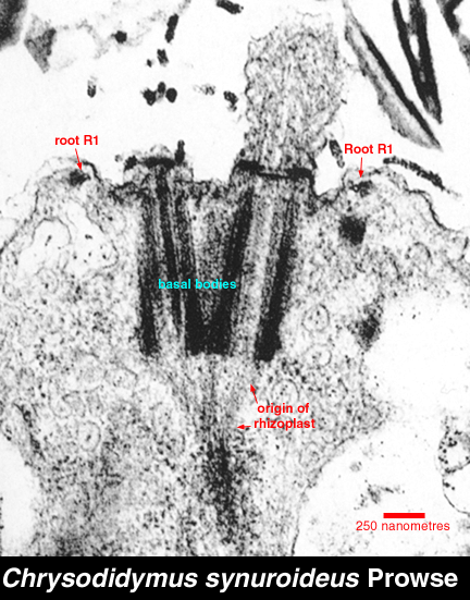
The two flagellar basal bodies are located at the anterior end of the
cell. They are nearly parallel to each other. Associated with the basal
bodies is a single microtubular root ("root R1") that forms a loop around
the anterior
end of the cell. From this microtubular root, cytoskeletal microtubules
descend towards the posterior end of the cell. There is also a striated
fiber (rhizoplast, System II fiber) that extends between the basal bodies
and the nucleus. This configuration of structures (the "kinetid" or
"flagellar apparatus") is characteristic for members of the
"synurophyte" golden algae.
|
 As in other golden algae, the mitochondrial cristae are tubular, and the
chloroplasts have three-thylakoid lamellae and are bounded by rough
endoplasmic reticulum (the "chloroplast endoplasmic reticulum"). In many
golden algae, the chloroplast endoplasmic reticulum is continuous with the
nuclear envelope, but this features is not apparent in
Chrysodidymus or other "synurophyte" algae. As with other
"synurophyte" algae, the scales are formed in the chloroplast
endoplasmic reticulum on the side of the chloroplast that faces the
cell surface. The large vacuole in
the posterior end of the cell is presumed to contain chrysolaminarin, the
storage polysaccharide.
As in other golden algae, the mitochondrial cristae are tubular, and the
chloroplasts have three-thylakoid lamellae and are bounded by rough
endoplasmic reticulum (the "chloroplast endoplasmic reticulum"). In many
golden algae, the chloroplast endoplasmic reticulum is continuous with the
nuclear envelope, but this features is not apparent in
Chrysodidymus or other "synurophyte" algae. As with other
"synurophyte" algae, the scales are formed in the chloroplast
endoplasmic reticulum on the side of the chloroplast that faces the
cell surface. The large vacuole in
the posterior end of the cell is presumed to contain chrysolaminarin, the
storage polysaccharide.
|
 There are a number of vacuoles in the anterior end of the cell. Their
function is unknown.
There are a number of vacuoles in the anterior end of the cell. Their
function is unknown.
|
Return to summary information


 As in other golden algae, the mitochondrial cristae are tubular, and the
chloroplasts have three-thylakoid lamellae and are bounded by rough
endoplasmic reticulum (the "chloroplast endoplasmic reticulum"). In many
golden algae, the chloroplast endoplasmic reticulum is continuous with the
nuclear envelope, but this features is not apparent in
Chrysodidymus or other "synurophyte" algae. As with other
"synurophyte" algae, the scales are formed in the chloroplast
endoplasmic reticulum on the side of the chloroplast that faces the
cell surface. The large vacuole in
the posterior end of the cell is presumed to contain chrysolaminarin, the
storage polysaccharide.
As in other golden algae, the mitochondrial cristae are tubular, and the
chloroplasts have three-thylakoid lamellae and are bounded by rough
endoplasmic reticulum (the "chloroplast endoplasmic reticulum"). In many
golden algae, the chloroplast endoplasmic reticulum is continuous with the
nuclear envelope, but this features is not apparent in
Chrysodidymus or other "synurophyte" algae. As with other
"synurophyte" algae, the scales are formed in the chloroplast
endoplasmic reticulum on the side of the chloroplast that faces the
cell surface. The large vacuole in
the posterior end of the cell is presumed to contain chrysolaminarin, the
storage polysaccharide.
 There are a number of vacuoles in the anterior end of the cell. Their
function is unknown.
There are a number of vacuoles in the anterior end of the cell. Their
function is unknown.